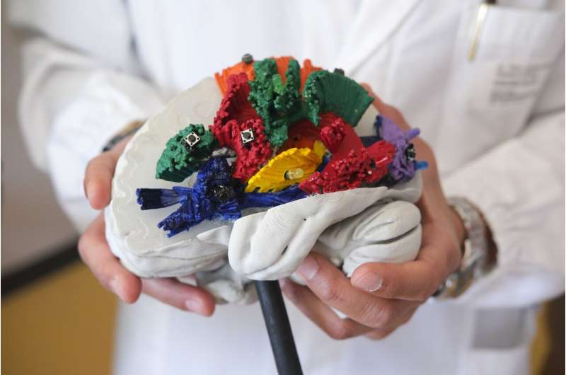
The published paper reports on the results of more than 5 years of collaboration and introduces a new instrument that the scientific community can use to accurately integrate ex-vivo dissection and in-vivo tractography data. These two complementary techniques have never been integrated so far into the study of human white matter connections, and this confirms a new research trend where multidisciplinary competencies converge, in this case clinical neuroscience and artificial intelligence.
The study opens new frontiers for neurosurgery in the treatment of brain tumors and the approach to degenerative neurological disorders, and in neuro-rehabilitation to harness the potential of brain plasticity.
BraDiPho was presented in a paper published in Nature Communications, with Laura Vavassori as first author. She is a doctoral student at the Center for Brain/Mind Sciences (Cimec) of the University of Trento.

