The research is published in the journal Advanced Science. The team was led by Professors Tae-Eun Park and Hyun-Wook Kang from the Department of Biomedical Engineering at UNIST, in collaboration with Professor Seung-Jae Myung from Seoul Asan Medical Center.
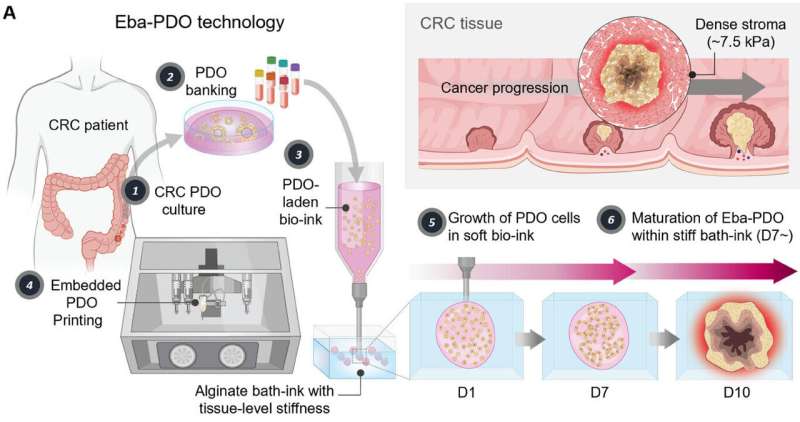
The team developed an artificial tumor tissue called the Embedded Bioprinting-enabled Arrayed PDOs (Eba-PDOs). This model accurately reproduces the high matrix stiffness and hypoxic conditions characteristic of real tumor tissues, particularly in colorectal cancer (CRC). By analyzing the morphology of these artificial tumors with AI, the team achieved 99% accuracy in predicting the expression of key prognostic gene markers, such as CEACAM5, in colorectal cancer.
Cancer cells proliferate rapidly, leading to dense, firm tissues with low oxygen levels—conditions that influence tumor growth and drug response. Traditional artificial tumor models, even those derived from patient cells, have struggled to faithfully mimic these environmental factors, resulting in distortions in observed growth patterns and treatment responses.