Until now, two methods—3D-PLI and ComSLI—have been used separately. Researchers from Jülich and Delft have developed a system that combines both methods to great advantage. They present their “Scattering Polarimeter” in a recent study published in the journal Scientific Reports.
Advantages of the combination
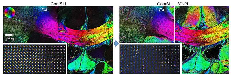
3D Polarized Light Imaging (3D-PLI) makes it possible to visualize the course of nerve fibers in entire histological brain sections with a resolution in the micrometer range using visible light. However, the method leaves uncertainties in pixels that contain intersecting nerve fibers. This problem can be solved with the help of Computational Scattered Light Imaging (ComSLI): The same brain slices are illuminated (now automated) from different angles and the transmitted (scattered) light is measured under normal incidence.
This produces light intensity profiles that reveal the underlying brain tissue structure and thus the crossing of nerve fibers. A combination of both methods in a single device would offer decisive advantages: faster measurements, pixel-by-pixel mapping and cross-validation of fiber courses.