With data from more than 39,000 participants from UK Biobank, this is one of the largest population-based studies to date exploring the relationship between heart anatomy and electrical activity. By combining 3D heart imaging with ECG data, the team created simplified digital twins of each participant’s heart.
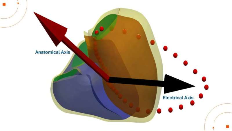
These personalized models allowed researchers to explore how the heart’s anatomical position, known as the anatomical axis, aligns with a spatial metric of electrical activity, or electrical axis. The study is published in the journal PLOS Computational Biology.
Digital twins are emerging as a powerful tool in cardiovascular research, enabling scientists to simulate and study the heart’s structure and function in unprecedented detail. In this study, they were key to revealing how natural variations in heart orientation, shaped by factors such as body mass index (BMI), sex, and hypertension, can significantly influence ECG readings.