During surgeries on brain vessels, for example, when treating an aneurysm or creating a bypass, there is a risk that the blood flow in brain tissue will temporarily stop. This can lead to a stroke. The risk of a stroke happening during aneurysm surgeries starts at around 8% and can rise to nearly 50% in complex cases. For brain tumors such as gliomas, the risk lies between 12.5% and 44%.
Pilot with 10 patients
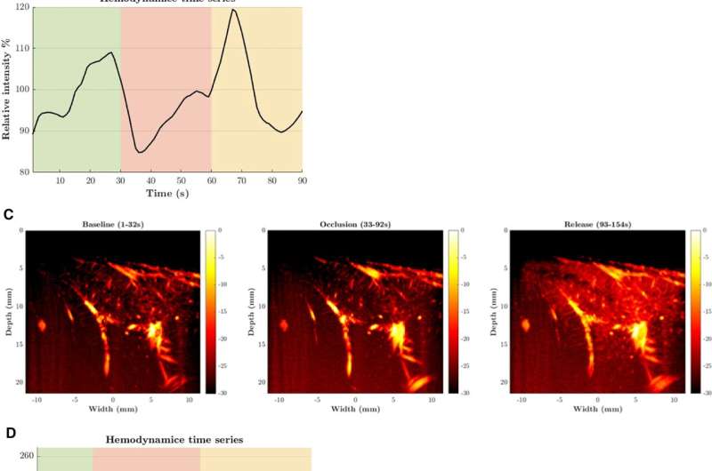
Until now, surgeons did not have a tool to directly observe a stroke during surgery. Thanks to a special type of ultrasound—Ultrafast Power Doppler Imaging (UPDI)—this has now changed. The technique visualizes the capillaries, measures the amount of blood in the brain tissue, and shows what happens during the procedure in real-time.
This allows surgeons to immediately see whether the brain tissue receives enough blood during the temporary closure of a blood vessel. The technique is fast, safe, and easy to use during surgery.