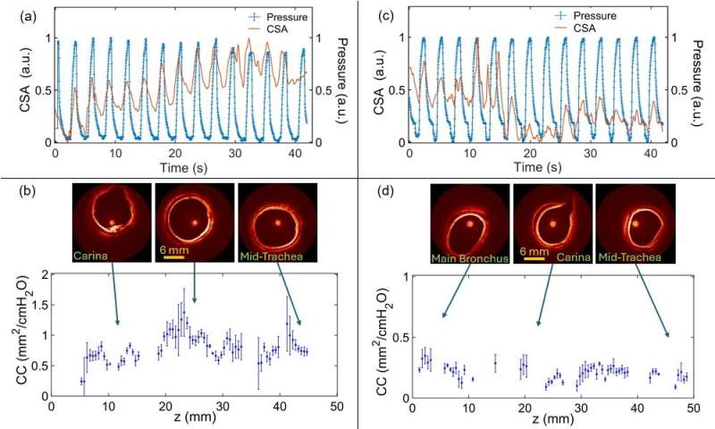
Airway wall elastography, performed with endoscopic optical coherence tomography (OCT), can detect subtle changes in how airway tissue deforms with breathing. By combining OCT’s high-resolution imaging with a pressure sensor, clinicians can calculate cross-sectional compliance—how much the airway expands or contracts under pressure. However, current methods require relatively long scan times, which can be impractical in clinical settings.
The new approach uses a “retrospective, respiratory-gated” 4D OCT scanning method. Instead of imaging the airway in sequence from one end to the other, the researchers move the scanning catheter in a sawtooth pattern along a 50 mm section. This motion allows each location to be captured at different points in the breathing cycle, both high and low pressure. Afterward, the data is sorted by position and phase of respiration to calculate compliance at each point, with a spatial resolution of 0.5 mm.