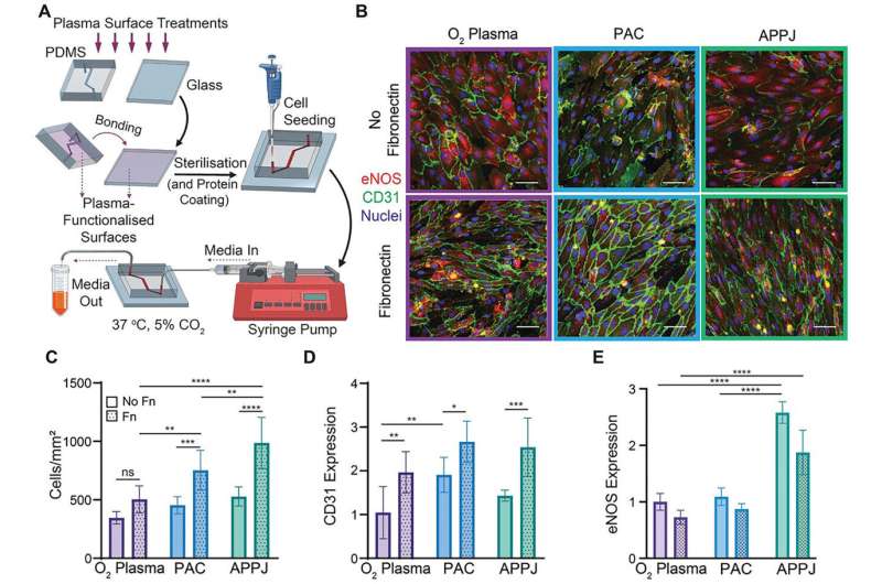
The innovative device mimics blood vessel damage due to high blood flow and inflammation, the first stage that leads to development of heart disease. The design offers a more accurate and detailed understanding of how and why blockages occur in specific locations in blood vessels. The development of the technology has been published in two journal articles.
“This is an incredible development because we took advantage of these microchips being made from a transparent material, and we mimicked the conditions of the coronary artery, that supplies blood to the heart muscle, and imaged them with a microscope to map the areas of cell damage which were similar to the locations of blockages in patients with heart disease,” says Associate Professor Anna Waterhouse from the Charles Perkins Center and the Sydney Nano Institute.
“If we use an animal model, we are unable to see changes at this level of detail in a living organism because you can’t see through the vessels.”

