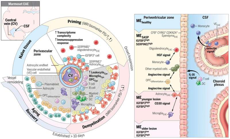
The researchers, led by postdoctoral fellow Jing-Ping Lin, Ph.D., and senior investigator Daniel S. Reich, M.D., Ph.D., both at NIH’s National Institute of Neurological Disorders and Stroke (NINDS), combined repeated MRI imaging with brain-tissue analysis, including gene expression, to track the onset and development of MS-like lesions.
They uncovered a new MRI signature that can help detect brain regions at risk for damage weeks before any visible lesions occur. They also identified “microenvironments” within affected brain tissue based on observed patterns of neural function, inflammation, immune and support cell responses, gene expression, and levels of damage and repair