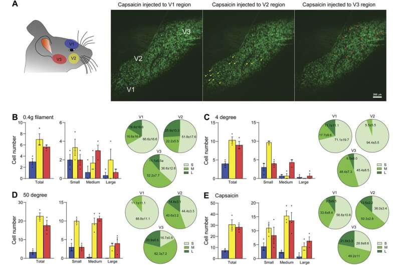
A study published in the Pain journal on December 2024 revealed a research team led by Yu Shin Kim, Ph.D., associate professor in the Department of Oral and Maxillofacial Surgery in the School of Dentistry at The University of Texas Health Science Center at San Antonio (UT Health San Antonio), observed for the first time the simultaneous activity of over 3,000 trigeminal ganglion (TG) neurons, which are cells clustered at the base of the brain that transmit information about sensations to the face, mouth and head.
“With our novel imaging technique and tools, we can see each individual neuron’s activity, pattern and dynamics as well as 3,000 neuronal populational ensemble, network pattern and activities in real time while we are giving different stimuli,” said Kim.
TMJ disorders are the second most common musculoskeletal disorder in the United States, affecting 8% to 12% of Americans. Current treatments often fall short, prompting researchers to explore the intricate nerve and vessel network surrounding TMJ.

