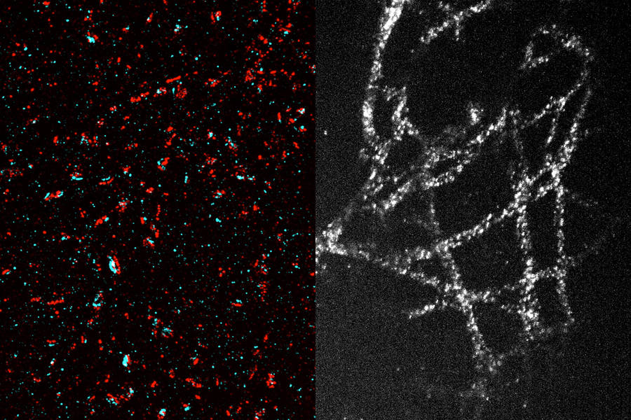
A classical way to image nanoscale structures in cells is with high-powered, expensive super-resolution microscopes. As an alternative, MIT researchers have developed a way to expand tissue before imaging it — a technique that allows them to achieve nanoscale resolution with a conventional light microscope.
In the newest version of this technique, the researchers have made it possible to expand tissue 20-fold in a single step. This simple, inexpensive method could pave the way for nearly any biology lab to perform nanoscale imaging.
“This democratizes imaging,” says Laura Kiessling, the Novartis Professor of Chemistry at MIT and a member of the Broad Institute of MIT and Harvard and MIT’s Koch Institute for Integrative Cancer Research. “Without this method, if you want to see things with a high resolution, you have to use very expensive microscopes. What this new technique allows you to do is see things that you couldn’t normally see with standard microscopes. It drives down the cost of imaging because you can see nanoscale things without the need for a specialized facility.”

