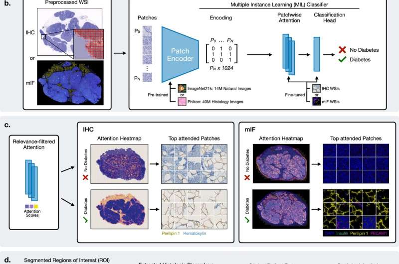
More than 500 million people worldwide live with type 2 diabetes. They often suffer from serious complications. Nevertheless, it has been difficult to draw reliable conclusions about a person’s glycemic status based on classic histopathological examinations. Many subtle morphological changes associated with impaired insulin secretion and beta cell dysfunction are barely visible to the naked eye.
To close this diagnostic gap, the research team created an extensive data set from pancreatic tissue sections from living donors. The samples were contrasted using chromogenic and multiplex immunofluorescent staining and then captured in high resolution using gigapixel microscopy.