The tool, called PICTURE (Pathology Image Characterization Tool with Uncertainty-aware Rapid Evaluations), distinguished with near-perfect accuracy between glioblastoma—the most common and aggressive brain tumor—and primary central nervous system lymphoma (PCNSL), a rarer cancer often mistaken for glioblastoma. While both can appear in the brain, glioblastoma arises from brain cells, whereas PCNSL develops from immune cells. Their similarities under the microscope often lead to misdiagnosis, with serious consequences for treatment.
The work is described Sept. 29 in Nature Communications. The AI model is publicly available for other scientists to use and build upon, the team said.
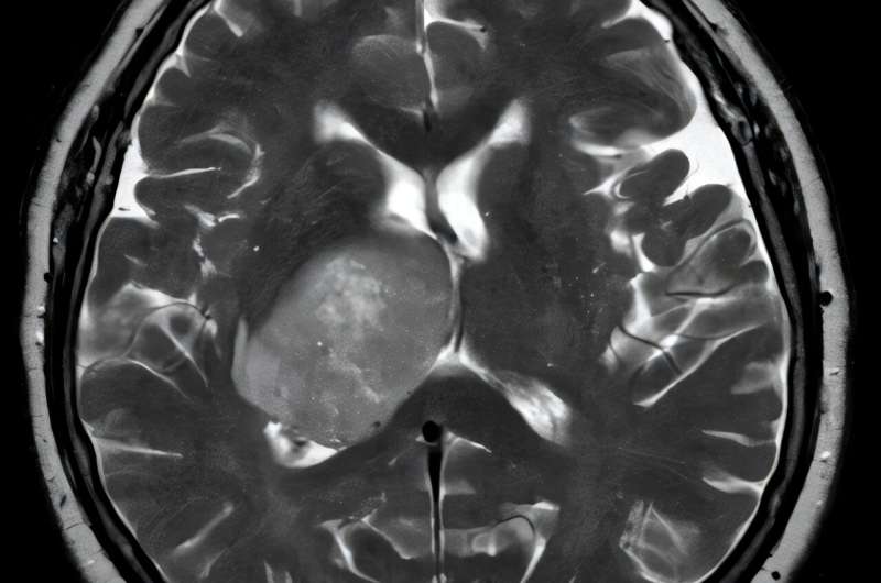
Correctly identifying look-alike tumors in the brain during surgery is one of the toughest diagnostic challenges in neuro-oncology, the researchers said. An accurate diagnosis while the patient is still in the operating room can help expedite critical treatment choices, such as whether to operate and remove the cancerous tissue—as should be done with glioblastoma—or leave it behind and opt for radiation and chemotherapy instead, the preferred therapy for PCNSL. Inaccurate or delayed diagnosis of cancers in the brain can lead to unnecessary surgery and delays in proper treatment.