
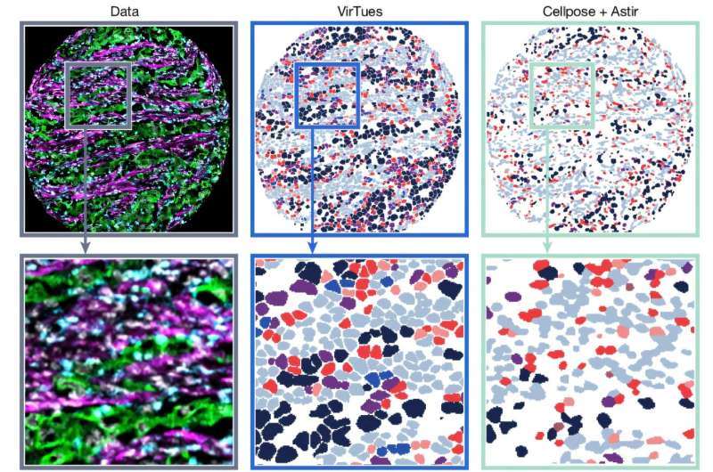
To address this challenge, Charlotte Bunne’s Artificial Intelligence in Molecular Medicine group at EPFL’s School of Computer and Communication Sciences and School of Life Sciences has developed Virtual Tissues (VirTues), a foundation model for tissue biology. The model learns from spatial proteomics data from different studies and cancer types and can be used to study biology across scales, from individual cells to whole tissue sections to patient outcomes. The results have been published in Nature.
Measuring dozens of proteins at once
“Which cells are present is only part of the picture,” Bunne says. “We also need to know where they are and how they interact.” Answering that across many patients is as much a computational problem as a biological one.