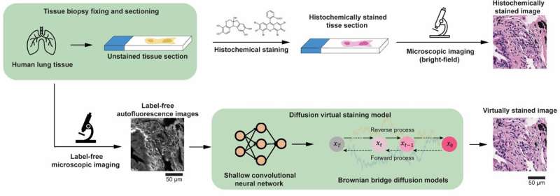
To overcome these limitations, “virtual staining” has emerged as a powerful computational tool that transforms images of unstained tissue into equivalents of these chemically stained samples, without the need for physical dyes or chemical procedures.
In a study published in Nature Communications, a team of researchers at the University of California, Los Angeles (UCLA) reported an AI tool that virtually stains unlabeled tissue samples at a resolution far exceeding that of the input image—without the use of any chemical dyes or staining.
By leveraging a cutting-edge diffusion model inspired by a Brownian bridge process, the method generates highly detailed and accurate microscopic images of tissue that digitally replace traditional histochemical staining, offering a non-destructive, cost-effective, and scalable alternative to digital pathology.