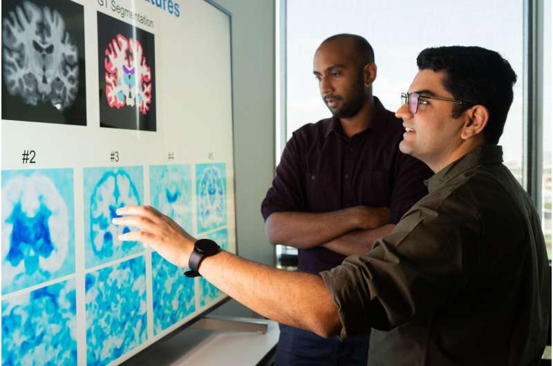
In the days before artificial intelligence (AI) and machine learning (ML), clinicians performed this crucial yet painstaking and time-consuming task by hand, but over the past decade, U-nets—a type of AI architecture specifically designed for medical image segmentation—has been the go-to instead. However, U-nets require large amounts of data and resources to be trained.
“For large and/or 3D images, these demands are costly,” said Kushal Vyas, a Rice electrical and computer engineering doctoral student and first author on a paper presented at the Medical Image Computing and Computer Assisted Intervention Society, or MICCAI.
“In this study, we proposed MetaSeg, a completely new way of performing image segmentation.”