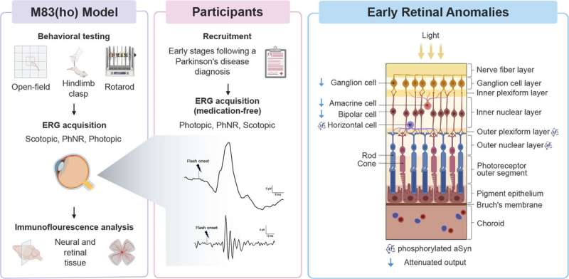
Parkinson’s disease is usually diagnosed when a person consults a doctor because of motor problems such as tremors. “By then, the disease has been present for several years and the affected neurons are already engaged in an irreversible degenerative process. That’s why it’s important to find biomarkers that detect Parkinson’s at an early stage of the disease,” explains study leader Martin Lévesque, professor at Université Laval’s Faculty of Medicine and researcher at CERVO Brain Research Center in Québec City.
“The retina is a direct extension of the central nervous system and, consequently, offers a non-invasive way of exploring the brain,” continues the researcher. “An unusual response of the retina to light stimuli could be indicative of a pathology affecting the brain.”
To explore this avenue, his research team recruited 20 people who had been diagnosed with Parkinson’s for less than 5 years. “We placed an electrode on each participant’s lower eyelid and recorded their retinal response to a series of flashes of different intensity, frequency and color. We did the same with people of the same age, but in good health. The results we obtained for people with Parkinson’s had a distinct signature from those in the control group,” explains Professor Lévesque.