“This work represents the first-ever subcellular resolution digital twin of a differentiated human primary cell, demonstrating how the eye is an ideal proving ground for developing methods that could be used more generally in biomedical research,” Kapil Bharti, Ph.D., scientific director at the NIH’s National Eye Institute (NEI).
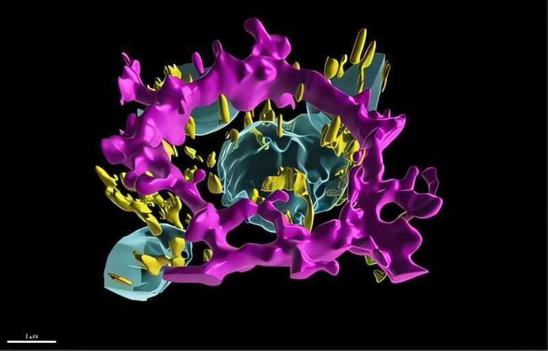
The researchers created a highly detailed, 3D data-driven digital twin of retinal pigment epithelial (RPE) cells, which perform vital recycling and supportive roles to light-sensing photoreceptors in the retina. In diseases such as AMD, RPE cells die, which eventually leads to the death of photoreceptor cells, causing loss of vision.
For RPE cells to do their multiple jobs properly, they require a top-to-bottom polarity: The cell’s “top” (apical) side faces photoreceptors, where they recycle worn out photoreceptor parts daily. The cell’s “bottom” (basal) side faces the blood supply where it brings in nutrients and oxygen and ships out waste.