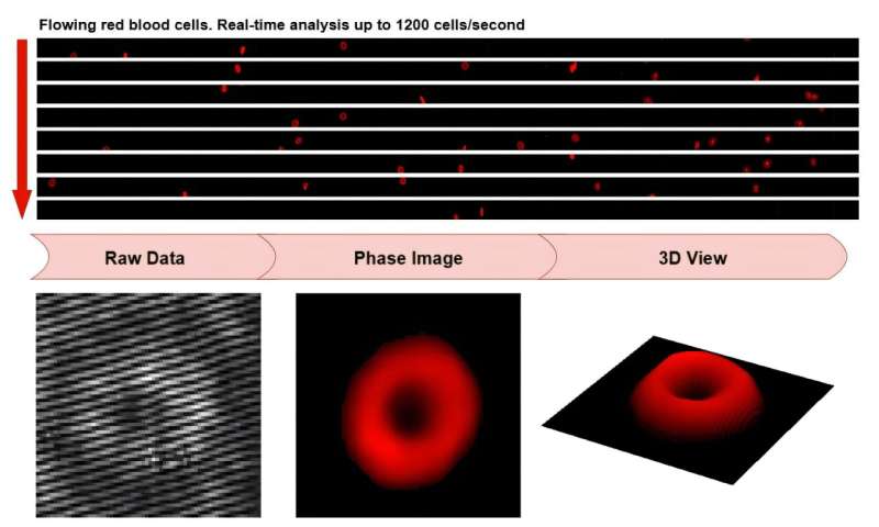
One promising technique, called quantitative phase microscopy (QPM), uses optical holography to measure the shape, thickness, and size of individual cells without any dyes or contrast agents, which provides quantitative 3D information to assist diagnostic decisions. For diseases that cause changes in cell morphology, such as sickle cell disease (SCD), this method enables a high-throughput diagnostic tool that can be used at the point of care.
High-throughput QPM systems image flowing red blood cells (RBCs) at a high frame rate, acquiring images of over 100,000 cells in under 3 minutes. After reconstruction, researchers can conduct statistical analysis of large numbers of cells, which allows quantification, for instance, of a patient’s SCD severity.
However, to analyze phase images, the QPM data must be digitally reconstructed. The large amount of data collected by high-throughput QPM can take several hours to process on a regular CPU, whereas fast, real-time processing typically relies on expensive, high-performance GPUs, making it difficult to balance diagnosis time and cost in clinical applications.