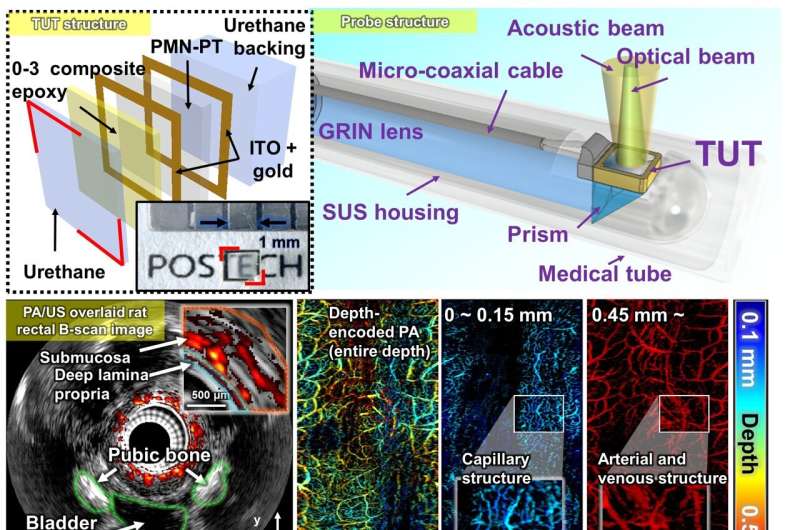
Endoscopic ultrasound is widely used in gastroenterology for cancer diagnosis, but it offers limited contrast in soft tissues and only provides structural information, ultimately reducing diagnostic sensitivity.
To address this, numerous studies have attempted to integrate photoacoustic technology with endoscopic ultrasound to provide more detailed information about tissue vasculature, thereby improving early cancer detection. However, achieving high-quality photoacoustic and ultrasound imaging simultaneously within an ultra-compact probe has proven challenging.
For obtaining high-resolution images, light and ultrasound must be aligned in the same direction. However, past efforts faced limitations in achieving this alignment. Ultimately, alignment could be achieved either by drilling a hole in the ultrasonic transducer to secure the light path or by tilting the optical system to align the two paths. Both approaches, however, come with trade-offs, often compromising the quality of either the ultrasound image or the photoacoustic image.

