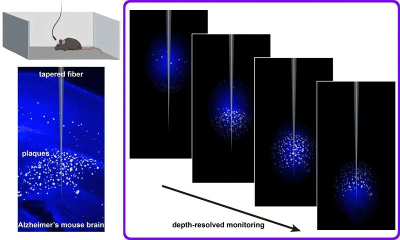
A new study, published in Neurophotonics, describes a fiber-optic method that can monitor plaque signals in living, freely moving mice, opening the door to more flexible testing of potential therapies.
The team, led by researchers at the University of Strathclyde and the Italian Institute of Technology, adapted fiber photometry—a technique commonly used to record neural activity—to detect amyloid plaques. Instead of relying on genetically encoded sensors, they used Methoxy-X04, a fluorescent dye that crosses the blood-brain barrier and binds specifically to amyloid fibrils.