
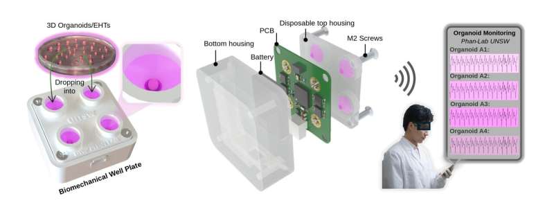
Cardiac organoids are 3D clusters of human heart cells grown in a laboratory that are used to evaluate the safety and efficacy of new drugs prior to clinical trials, as well as study disease. While they don’t replicate the full structure of a human heart, they mimic key behaviors, especially how heart muscles contract when drugs are administered.
They are increasingly seen as a powerful alternative to animal models, which often fail to fully capture how human biology works.
Current ways of monitoring whether these cardiac organoids are working properly often rely on optical imaging, essentially filming the organoids under microscopes and analyzing the footage. This process is time-consuming, difficult to scale and can disrupt the delicate environment the tissues need to survive.