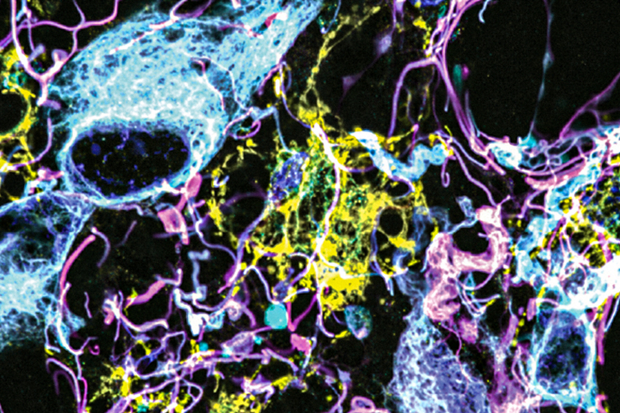
Using a novel microscopy technique, MIT and Brigham and Women’s Hospital/Harvard Medical School researchers have imaged human brain tissue in greater detail than ever before, revealing cells and structures that were not previously visible.
Among their findings, the researchers discovered that some “low-grade” brain tumors contain more putative aggressive tumor cells than expected, suggesting that some of these tumors may be more aggressive than previously thought.
The researchers hope that this technique could eventually be deployed to diagnose tumors, generate more accurate prognoses, and help doctors choose treatments.
“We’re starting to see how important the interactions of neurons and synapses with the surrounding brain are to the growth and progression of tumors. A lot of those things we really couldn’t see with conventional tools, but now we have a tool to look at those tissues at the nanoscale and try to understand these interactions,” says Pablo Valdes, a former MIT postdoc who is now an assistant professor of neuroscience at the University of Texas Medical Branch and the lead author of the study.