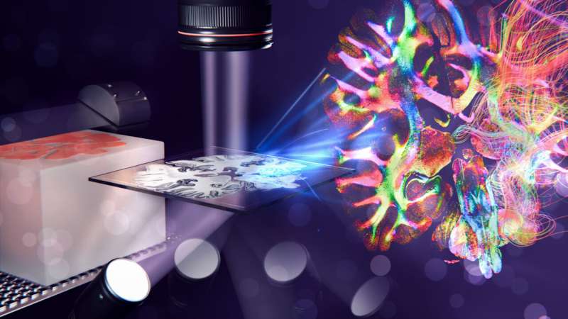
Researchers from Delft, Stanford, Jülich, and Rotterdam achieved a milestone: using a technique called ComSLI, they enable fiber mapping inside any tissue section with micrometer precision. The findings are published in Nature Communications.
Studying the network of neurons in the brain helps to understand brain diseases such as Alzheimer’s or Parkinson’s disease. To analyze the detailed anatomy under a microscope, the tissue is often embedded in paraffin wax and cut into micrometer-thin slices.
These so-called formalin-fixed paraffin-embedded (FFPE) sections are the gold standard for studying healthy or diseased tissues. However, current microscopy techniques cannot accurately map large nerve fiber networks in these FFPE sections.