The new approach, reported in the Journal of Medical Imaging, combines light-emitting diodes (LEDs) with hyperspectral imaging technology to create detailed maps of tissue properties that are invisible to conventional endoscopic cameras.
Unlike standard endoscopy, which captures images using broad red, green, and blue color channels, hyperspectral imaging records data across numerous narrow wavelength bands, including light beyond the visible spectrum. This allows the system to detect biochemical changes in cancerous tissue that produce distinct spectral signatures.
Testing the LED array concept
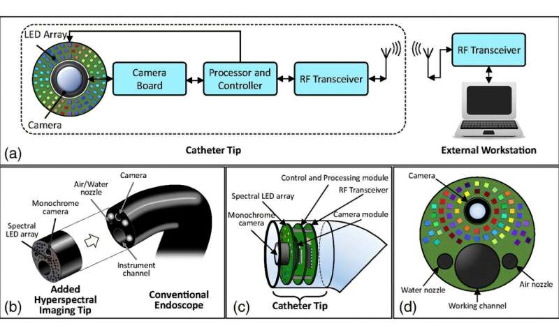
The research team led by Dr. Baowei Fei, Professor and Cecil H. and Ida Green Chair in Systems Biology Science at the University of Texas at Dallas (UT Dallas) Quantitative BioImaging Laboratory, designed and tested a prototype system built around an array of 18 LEDs, each emitting light at different wavelengths ranging from 405 nanometers to 910 nanometers. The system uses a monochrome camera to capture images as each LED illuminates the target tissue in sequence, building up a complete hyperspectral dataset.