A research team at Okayama University, Japan, led by Professor Masazumi Fujiwara and his Ph.D. student Sara Mandic, in collaboration with Professor Ron M. A. Heeren of Maastricht University, Netherlands, has developed a new microfluidics-based workflow that enables high-resolution, 3D lipid imaging in C. elegans. Their findings were published in the journal Scientific Reports on July 8, 2025.
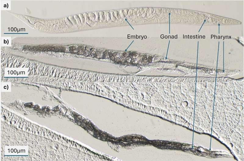
The team combined matrix-assisted laser desorption/ionization mass-spectrometry imaging (MALDI-MSI) with conventional lipid staining techniques to map both the identity and location of lipid molecules inside the worm. To preserve internal structures, young adult nematodes were aligned and immobilized on a custom-designed microfluidic chip, embedded in a gelatin–carboxymethyl cellulose mixture, sectioned using a cryotome and analyzed through MALDI-MSI. Each section was also subjected to Oil Red O staining, which highlights neutral fats, to confirm and complement the imaging results.