The neural network model developed in the study outperformed all previous models in the classification of tissue microscopy samples. The research is published in the journal Heliyon.
“Based on our study, the developed model is able to identify all tissue categories relevant for cancer identification, with an accuracy of 96.74%,” Fabi Prezja, the researcher responsible for the design of the method, says.
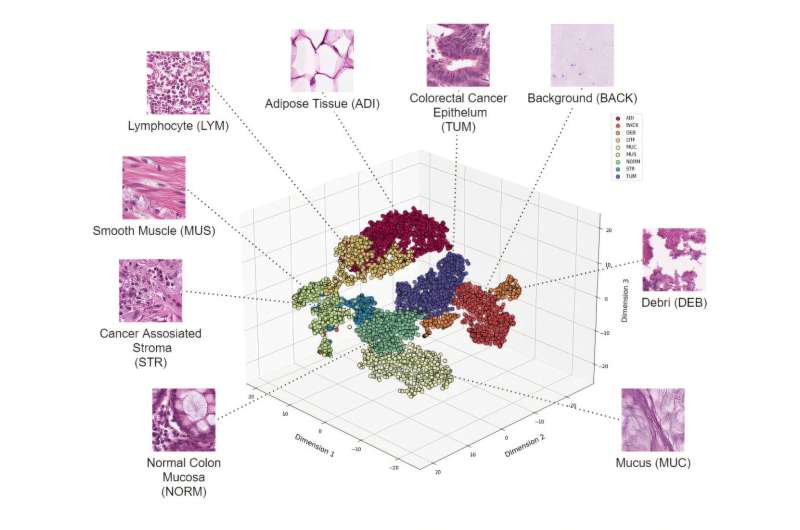
In practice, the tissue analysis involves a pathologist looking through the scanned digital microscopy slides, prepared from the patient’s intestine sample, and marks, point by point, for example, where the cancerous and related tissues are visible.