The work, published in Biophotonics Discovery, presents the first demonstration of this method in living human eyes.
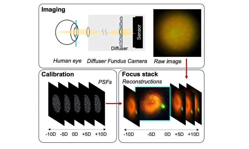
To build the system, the team modified a commercial fundus camera, replacing part of its optics with a holographic diffuser and sensitive digital sensor. They then calibrated the system by recording how the diffuser blurs light with different amounts of refractive error. Once calibrated, the camera could record images of the retina in model eyes and digitally sharpen them to the correct focus using software.
In tests with volunteers, the device successfully captured color images of the retina, showing features such as the optic disk, blood vessels, and macula. Importantly, the images could be refocused across a wide range of refractive errors—more than ten diopters—without moving any optical components. The system produced consistent resolution of about 7 to 10 line pairs per millimeter, which is lower than that of a conventional camera but remained stable for eyes with refractive errors, all with fixed optics and no prior focusing.