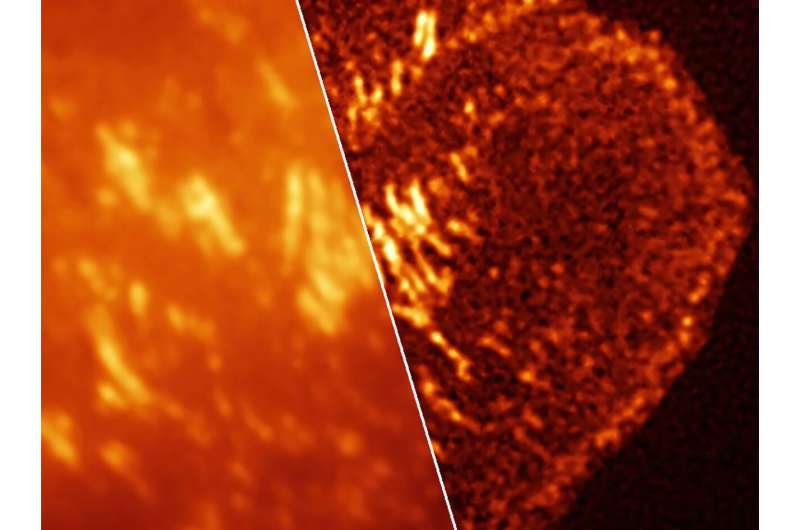
Researchers use a technique called super‑resolution microscopy to see tiny structures inside cells that are normally invisible with standard light microscopes. While powerful, this approach can be difficult to use in living cells. Traditional methods often require special light patterns or harsh chemicals to force fluorescent tags to turn on and off, which can damage cells and discourage many labs from using the technology.
The newly developed dyes solve this problem in a simpler way. They blink on and off naturally, without the need for intense light or added chemicals. That makes it easier for scientists to localize individual molecules inside living cells using standard lab equipment. It also makes them well-suited for SOFI—super-resolution optical fluctuation imaging—which uses changes in fluorescence intensity to build high-resolution images much faster than localizing individual molecules.