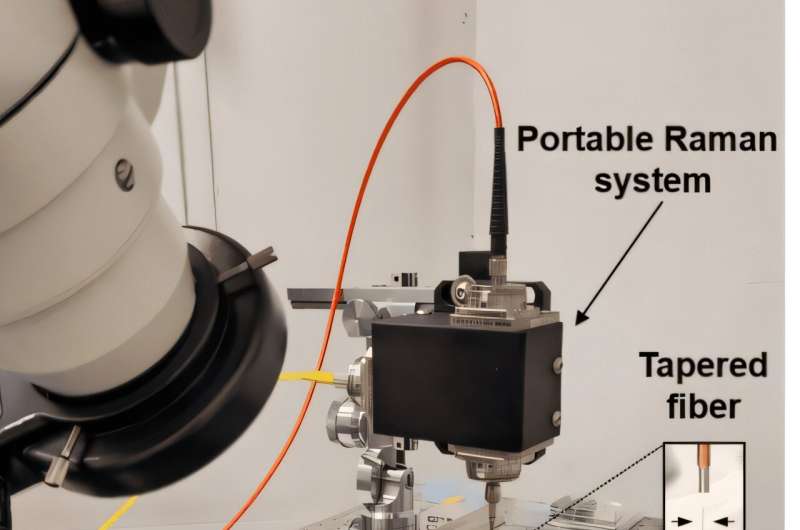
The authors describe the new technique as a “molecular flashlight” because it provides information about the chemical composition of nerve tissue by illuminating it. This makes it possible to analyze molecular changes caused by tumors, whether primary or metastatic, as well as injuries such as a traumatic brain injury.
The molecular flashlight is a probe less than 1 mm thick, with a tip just one micron wide—about one thousandth of a millimeter—and invisible to the naked eye. It can be inserted deep into the brain without causing damage (for comparison, a human hair measures between 30 and 50 microns in diameter).
This flashlight-probe is not yet ready to be tested in patients, and for now it is primarily a “promising” research tool in animal models that allows “monitoring molecular changes caused by traumatic brain injury, as well as detecting diagnostic markers of brain metastasis with high accuracy,” explain the authors of the paper.

