
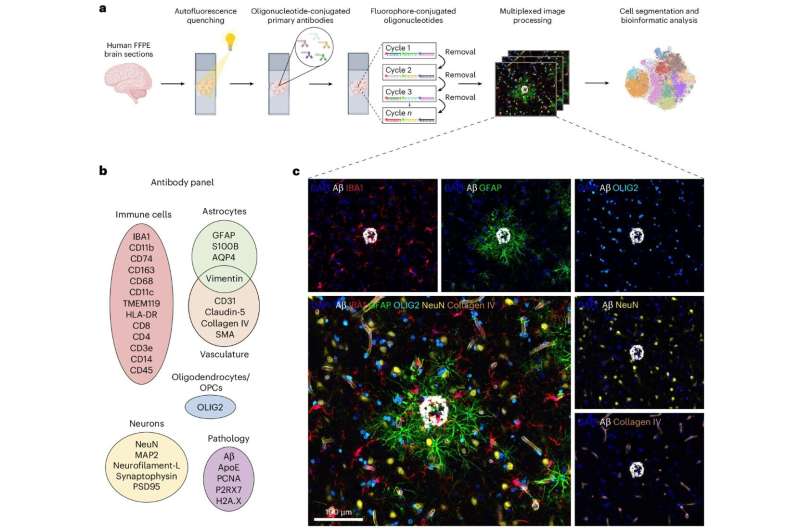
A newly developed microscopy technique allows, for the first time, the visualization of more than 30 protein markers simultaneously in the human brain and uses bioinformatics to analyze their spatial relationships.
The development could, in the future, bring about lasting changes not only in Alzheimer’s research but also in the investigation of tumors and other diseases. The method, known as CODEX-CNS, can visualize different cell types simultaneously, investigate their functional properties and, furthermore, analyze the spatial relationships between cells. The research findings have just been published in Nature Neuroscience.