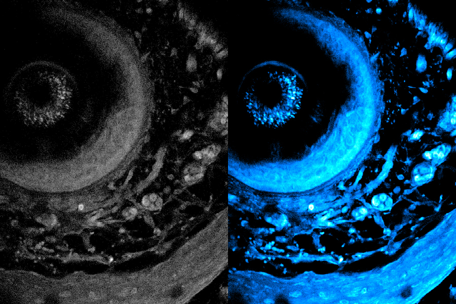
Metabolic imaging is a noninvasive method that enables clinicians and scientists to study living cells using laser light, which can help them assess disease progression and treatment responses.
But light scatters when it shines into biological tissue, limiting how deep it can penetrate and hampering the resolution of captured images.
Now, MIT researchers have developed a new technique that more than doubles the usual depth limit of metabolic imaging. Their method also boosts imaging speeds, yielding richer and more detailed images.
This new technique does not require tissue to be preprocessed, such as by cutting it or staining it with dyes. Instead, a specialized laser illuminates deep into the tissue, causing certain intrinsic molecules within the cells and tissues to emit light. This eliminates the need to alter the tissue, providing a more natural and accurate representation of its structure and function.

