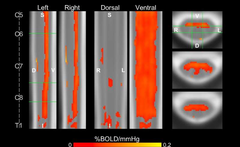
“We’ve taken MRI methods that we’ve firmly established in the brain, and with a lot of attention to detail and many small improvements, we’ve made this critical jump to use them in the spinal cord, and I find that pretty exciting,” said Molly Bright, DPhil, assistant professor of Physical Therapy and Human Movement Sciences and of Biomedical Engineering in the McCormick School of Engineering, who was senior author of the study.
Impaired vascular function in the spinal cord contributes to many neurological diseases, including traumatic spinal cord injury and degenerative cervical myelopathy—a condition where age-related damage to the spinal disks compresses the spinal cord, which can cause progressive weakness, numbness and difficulty with coordination.

