Histopathology currently relies on slicing tissue samples, extracted in a biopsy, into extremely thin sections, staining them with dyes and examining each slice under a microscope. These 2D images are stacked on top of each other to create a 3D image and then interpreted by specialists to identify disease.
The process is time-consuming, costly and destructive, meaning crucial follow-up tests that could help confirm the correct diagnosis often cannot be performed.
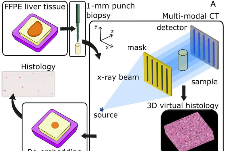
The newly developed, non-destructive approach, described in a new paper published in Proceedings of the National Academy of Sciences, uses a compact X-ray microscope based on standard anode X-ray source technology, the same used in most hospitals today. It is roughly the size of a small laboratory instrument and can generate high-resolution 3D maps of intact tissue samples, significantly cutting down on time and cost.