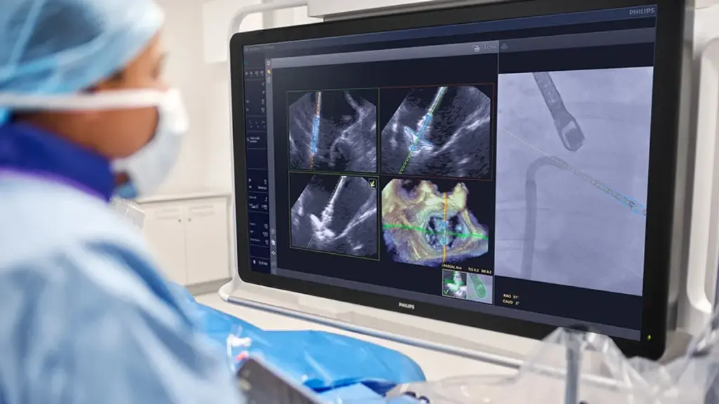
Physicians use X-ray fluoroscopy and 3D transesophageal echocardiography to visualize the heart and device during TEER procedures. However, the technologies create two separate images. The surgical team must interpret where the device is in the body based on the two images, potentially leading to miscommunication and suboptimal device placement.
Philips designed its software to combine the outputs of the two imaging modalities to continuously show the location of the device in the body. By visualizing the device in the ultrasound image, the companies aim to enable surgeons to more consistently place the repair system in the optimal location.