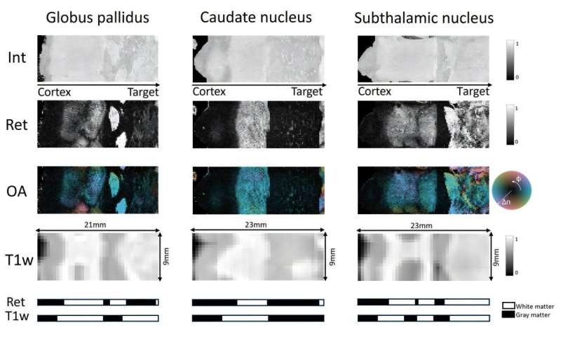
Success depends heavily on placing these electrodes with millimeter-level accuracy. However, current imaging tools like magnetic resonance imaging (MRI) often fall short of clearly identifying small, deep brain structures, making precise targeting difficult.
A recent study published in Neurophotonics explores a promising solution: catheter-based polarization-sensitive optical coherence tomography (PS-OCT).
PS-OCT is an optical imaging technique that uses polarized light to detect subtle structural differences in tissue. Unlike MRI, which provides millimeter-scale resolution, PS-OCT can visualize brain structures at the micrometer level. This allows it to detect fine details in white matter fiber tracts—bundles of nerve fibers that are crucial landmarks for DBS targeting.

