
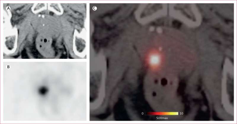
In an analysis of 194 patients, the method detected 91% of clinically relevant prostate cancers. The findings of the research team, led by Marcus Hacker, head of the Division of Nuclear Medicine at the Department of Biomedical Imaging and Image-guided Therapy, were published in the inaugural issue of the new journal The Lancet Medical Imaging & Theranostics.
PSMA-PET/CT combines two imaging techniques. In positron emission tomography (PET), a small amount of a radioactively labeled substance is administered, which binds specifically to the so-called prostate-specific membrane antigen (PSMA). This protein is particularly abundant on prostate cancer cells. This enables suspicious areas to be visualized. The computed tomography (CT) scan, carried out simultaneously, provides precise images of the body’s structures and enables these areas to be anatomically localized.