Durotomy is a common neurosurgical complication involving a tear in the dura mater, the protective membrane surrounding the brain and spinal cord. Damage can cause cerebrospinal fluid (CSF) leakage, leading to delayed healing, headaches, and infection, making a reliable watertight dural closure essential.
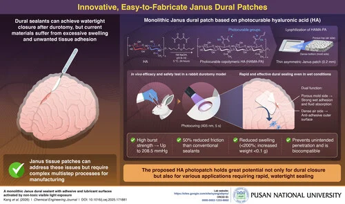
Tissue adhesives are being explored as more robust alternatives to suturing for dural closure, but many glue-based sealants swell excessively, causing mass effect, unwanted adhesion, and postoperative complications. Janus tissue patches, with one adhesive surface and one anti-adhesive surface, offer a potential solution; however, most rely on multiple materials and complex fabrication processes, limiting their clinical practicality.
In a breakthrough study, a research team from South Korea led by Professor Seung Yun Yang from the Department of Biomaterials Science at Pusan National University has developed an innovative light-responsive, monolithic Janus dural patch using photocurable hyaluronic acid (HA) through a simple approach. “Made from natural biopolymer hyaluronic acid, our dural patch provides strong wet adhesion, along with a lubricating surface that prevents unwanted tissue adhesion, after exposure to non-toxic visible light,” explains Prof. Yang. Their study was made available online on December 16, 2025, and published in Volume 527 of Chemical Engineering Journal on January 01, 2026.