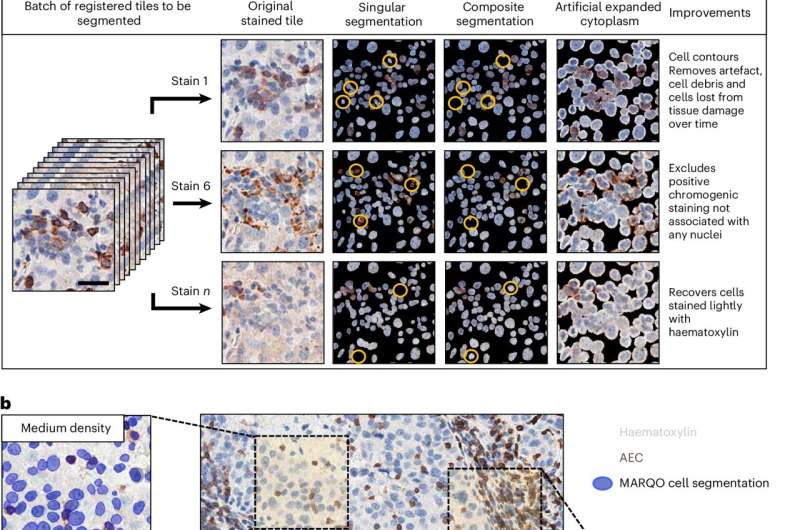
Developed by a team led by Sacha Gnjatic, Ph.D., Professor of Immunology and Immunotherapy at the Icahn School of Medicine, MARQO streamlines the complex task of analyzing immunohistochemistry (IHC) and immunofluorescence (IF) images, which are produced via staining methods commonly used to detect immune cells and other biomarkers in cancerous tissues.
When someone has cancer, pathologists inspect stained tissue sections under the microscope to see which cells are present and how they are arranged. Doing this by hand is labor-intensive and usually limited to small areas of the sample.
MARQO tackles this challenge in three key ways: First, while other tools can process entire images, they often require users to chop slides into patches or rely on costly computing clusters. MARQO keeps slides intact and finishes the job in minutes rather than hours, even on standard graphics processing units.

