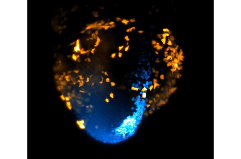
For the study, published in The EMBO Journal, the team used a technique called advanced light-sheet microscopy on a specially engineered mouse model. This is a method where a thin sheet of light is used to illuminate and take detailed pictures of tiny samples, creating clear 3D images without causing any damage to living tissue.
By doing this, they were able to track individual cells as they moved and divided over the course of two days—from a critical stage of development known as gastrulation through to the point where the primitive heart begins to take shape. This allowed the researchers to identify the cellular origins of the heart.
Gastrulation is the process by which cells begin to specialize and organize into the body’s primary structures, including the heart. In humans, this occurs during the second week of pregnancy.