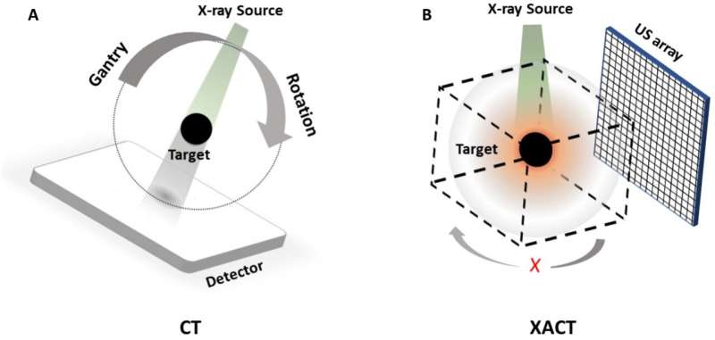
Computed tomography (CT) has long been a cornerstone of modern imaging, providing detailed 3D insights into the human body and other materials. However, conventional CT requires hundreds of X-ray projections from multiple angles, exposing patients to significant radiation doses and relying on large, immobile systems.
To address this issue, researchers from UC Irvine’s Departments of Radiological Sciences and Biomedical Engineering recently published a study in the journal Science Advances in which they introduce a technology that achieves 3D imaging with a single X-ray projection called X-ray–Induced Acoustic Computed Tomography (XACT).
A new paradigm in imaging
“In XACT, the generated sound waves by X-rays change the way X-ray imaging works, converting X-rays to ultrasound. X-rays typically travel in straight lines, so one projection only provides 2D information. However, X-ray-induced acoustic signals propagate in three dimensions, allowing for 3D imaging with a single projection,” said Shawn Xiang, Ph.D., the study’s corresponding author and an associate professor at UCI’s Departments of Radiological Sciences and Biomedical Engineering.

