The research is published in the journal Science. The team included researchers from Amsterdam UMC, VU LaserLab, the Netherlands Institute for Neuroscience and the University of Edinburgh.
The research team used advanced microscopy techniques and different models—from zebrafish and mouse models to human brain tissue—to research the formation of this damage.
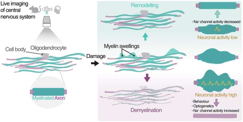
This led to the discovery that myelin swellings have a dynamic character: they can not only grow, but also shrink and even recover completely. It also turns out that the activity of the underlying nerve fiber plays an important role; more activity of the nerve fiber leads to more and bigger swellings, while less activity allows for possible recovery.