Supercharged ordinary clinical device gets a better look at the back of the eye

Scientists at the National Institutes of Health (NIH) have leveraged artificial intelligence to transform a device designed to see tissues in the back of the eye into one sharp enough to make out individual cells. The technique provides imaging resolution that rivals the most advanced devices available and is cheaper, faster, and doesn't require specialized equipment or expertise. The strategy has implications for early detection of disease and for the monitoring of treatment response by making what was once invisible now visible.

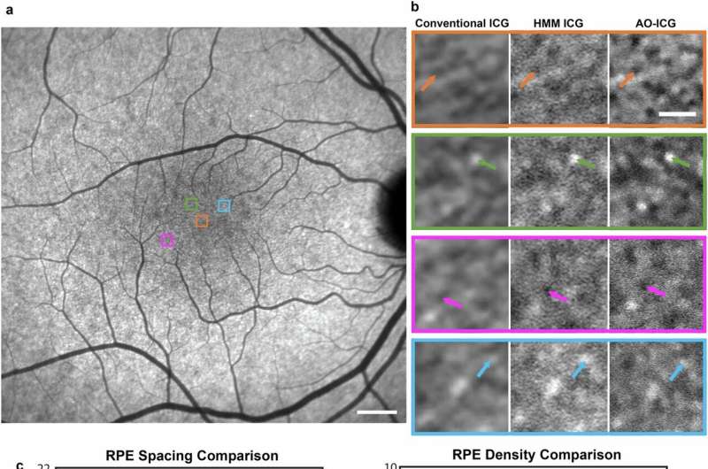
“AI potentially puts next-generation imaging in the hands of standard eye clinics. It’s like adding a high-resolution lens to a basic camera,” said Johnny Tam, Ph.D., investigator at NIH’s National Eye Institute and senior author of the study report, which was published in Communications Medicine.

Imaging devices, known as ophthalmoscopes, are widely used to examine the light-sensing retina in the back of the eye. A scanning laser ophthalmoscope is standard in eye clinics, but its resolution can only make out structures at the tissue level—things such as lesions, blood vessels, and the optic nerve head.

Sign up for Blog Updates