Blood microcirculation is a complex network that transports blood to tissues and organs through tiny blood vessels. When this mechanism functions properly, cells receive the oxygen and nutrients they need to stay healthy, while metabolic waste products are efficiently removed.
Any alteration in this network, whether structural or functional, can have serious clinical consequences, including heart failure, kidney failure and various chronic diseases. However, there is currently no imaging method that can visualize microcirculation and assess the integrity of the entire circulatory system, from the large arteries to the finest arterioles, at the level of the whole organ.
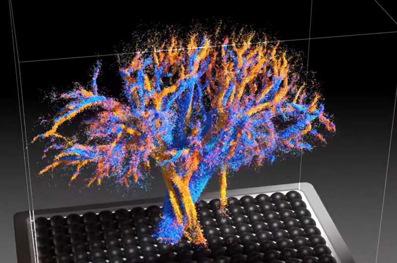
With this issue in mind, the Physics for Medicine Institute (Inserm/ESPCI Paris-PSL/CNRS) research team has developed the first tool capable of producing these images. It is a new type of ultrasound probe, developed as part of Nabil Haidour’s thesis work, under the supervision of Clément Papadacci (Inserm researcher).