
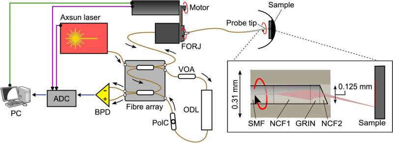
OCT is already an important tool for the diagnosis of eye disease due to its unique ability to provide high-resolution, cross-sectional images through the retina. Imaging is normally performed from outside of the eye, with images captured by rapidly scanning a low-power laser beam through the pupil. But for some surgical procedures, it may be beneficial to have a very small OCT endoscope inserted into the eye itself.
Most compact OCT endoscopes are side-viewing; when introduced into tubular structures such as blood vessels, they generate images of the vessel walls. Making an endoscope that is forward-viewing without increasing the diameter is much more difficult, but this is exactly what is needed for use inside the eye.