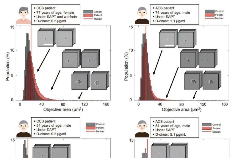
If you’ve ever cut yourself, you’ve seen platelets in action—these tiny blood cells are like emergency repair workers, rushing to plug the damage and stop bleeding. But sometimes, they overreact. In people with heart disease, they can form dangerous clots inside arteries, leading to heart attacks or strokes.
“Platelets play a crucial role in heart disease, especially in CAD, because they are directly involved in forming blood clots,” explained Dr. Kazutoshi Hirose, an assistant professor at the University of Tokyo Hospital and lead author of the study.
“To prevent dangerous clots, patients with CAD are often treated with antiplatelet drugs. However, it’s still challenging to accurately evaluate how well these drugs are working in each individual, which makes monitoring platelet activity an important goal for both doctors and researchers.”

