How does information flow through the brain? To understand this, researchers map the brain at every scale, from small networks of cells to the entire nervous system. This provides insight into how our brains work and how connections between cells may become disrupted in disease.
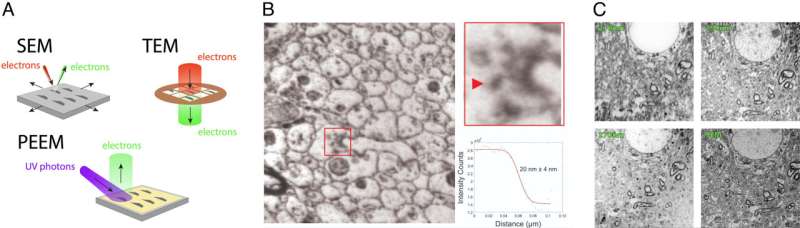
The research group led by Professor Sense Jan van der Molen uses a microscope that reveals how a brain structure is built. It can do so down to the level of a synapse, the tiny junction through which one neuron communicates with another cell.
Introducing the PEEM microscope in brain research
In a joint Chicago-Leiden project, Photoemission Electron Microscopy (PEEM) is introduced as a new tool for imaging brain tissue. In PEEM, the photo-electric effect is used to create images of materials of choice. Now, the researchers have applied it to image ultra-thin brain slices, taken consecutively from the same mouse brain.