Blog

- ARTIFICIAL INTELLIGENCE

AI helps scientists correct mistakes in medical studies

Randomized, controlled clinical trials are crucial for telling whether a new treatment is safe and effective. But often scientists don’t fully report the details of their trials in a way that allows other researchers to gauge how well they designed and conducted those studies.

Read More »

An LLM that can process and display transmitted cardiac data in real time

In addition to linguistic prompts, large language models can also understand, interpret, and adapt their responses to heart frequency data. Dr. Morris Gellisch, previously of Ruhr University Bochum, Germany, and now at University of Zurich, Switzerland, and Boris Burr from Ruhr University Bochum have developed a technical interface through which the physiological data can be transmitted to the language model in real time.

Read More »

Automated algorithm can detect cancer in blood samples in as little as 10 minutes

When cancer spreads, tiny amounts of cells can break away from tumors and circulate in the bloodstream. A liquid biopsy is a means to detect the presence of cancer by detecting these cancer cells floating in blood samples. However, current state-of-the-art methods have necessitated trained specialists to comb through and review images of thousands of cells out of potentially millions of cells on a slide over a period of many hours.

Read More »

AI-assisted growth prediction advances orthodontics

Orthodontic treatment is most effective when timed to coincide with a child’s growth peak. Traditionally, clinicians estimate growth by examining X-ray images of the cervical vertebrae—the neck bones visible in routine dental radiographs. However, this process requires careful manual annotation of specific points on the bones, a task that is both time-consuming and prone to variation between observers.

Read More »

AI tool could make medical imaging process 90% more efficient

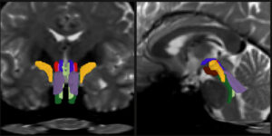
When doctors analyze a medical scan of an organ or area in the body, each part of the image has to be assigned an anatomical label. If the brain is under scrutiny, for instance, its different parts have to be labeled as such, pixel by pixel: cerebral cortex, brain stem, cerebellum, etc. The process, called medical image segmentation, guides diagnosis, surgery planning and research.

Read More »